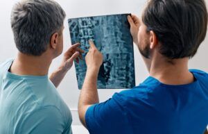
In the world of orthopedic care, accurate diagnosis is crucial to effective treatment. The specialists at Spine, Neck, & Back Specialists, led by Dr. Jay Reidler, serve the communities of Bloomfield, Englewood, and Union City, NJ, with a commitment to precision in diagnosis. Diagnostic imaging is key in identifying musculoskeletal conditions, from simple fractures to complex joint and spinal issues. This blog explores the three most commonly used imaging techniques in orthopedics: X-rays, MRI, and CT scans. Understanding the strengths and applications of each can help you make informed decisions about your health.
X-Rays: The First Line of Diagnostic Imaging
X-rays are the most commonly used diagnostic tool in orthopedic care, offering a quick and reliable way to visualize bones. By passing a small amount of radiation through the body, X-rays create images that allow doctors to assess bone structures and detect abnormalities.
What X-Rays Are Best For:
- Fractures: X-rays are excellent for detecting breaks or cracks in bones. They can determine the severity of a fracture and help guide treatment decisions.
- Bone Density: Conditions like osteoporosis, which result in weakened bones, can be assessed using X-rays to measure bone density.
- Joint Conditions: Arthritis, including osteoarthritis and rheumatoid arthritis, can be diagnosed by examining joint spaces and bone changes on an X-ray.
Advantages of X-Rays:
- Quick and Accessible: X-rays are readily available at most medical facilities, making them a convenient first step in diagnosis.
- Non-Invasive: The procedure is painless and typically takes only a few minutes.
- Cost-Effective: X-rays are generally more affordable than other imaging options, making them accessible to a wide range of patients.
Limitations of X-Rays: While X-rays are excellent for visualizing bones, they are less effective for evaluating soft tissues like muscles, ligaments, and tendons. For a more comprehensive view, additional imaging may be necessary.
MRI: Detailed Imaging of Soft Tissues
Magnetic Resonance Imaging (MRI) is a powerful diagnostic tool that uses strong magnetic fields and radio waves to produce detailed images of soft tissues. Unlike X-rays, MRI does not involve radiation, making it a preferred option for examining muscles, tendons, ligaments, and spinal discs.
What MRI Is Best For:
- Soft Tissue Injuries: MRI is ideal for diagnosing injuries to muscles, ligaments, and tendons. It can detect tears, strains, and inflammation with high precision.
- Spinal Conditions: Herniated discs, spinal stenosis, and nerve compression are best evaluated using MRI, as it provides clear images of the spine’s structure.
- Joint Evaluation: MRI is commonly used to assess complex joint conditions, including cartilage damage, meniscus tears, and ligament injuries.
Advantages of MRI:
- No Radiation: MRI avoids the use of ionizing radiation, making it a safer option for repeated scans or for vulnerable populations like pregnant women.
- High Resolution: The level of detail provided by MRI images is unmatched, especially for soft tissues.
- Versatile: MRI can capture images in multiple planes, offering a comprehensive view of the affected area.
Limitations of MRI: MRI scans can be time-consuming, often taking 30 to 60 minutes. Additionally, patients with certain metal implants or devices may not be candidates for MRI due to the magnetic fields involved.
CT Scans: Cross-Sectional Imaging for Detailed Analysis
Computed Tomography (CT) scans combine X-ray technology with computer processing to create cross-sectional images of the body. These detailed slices provide an in-depth view of bone structures, organs, and soft tissues, making CT an invaluable tool for diagnosing complex orthopedic conditions.
What CT Scans Are Best For:
- Complex Fractures: CT scans are particularly useful for evaluating fractures that are difficult to visualize with standard X-rays, such as those involving joints or the spine.
- Bone Abnormalities: Bone tumors, infections, and deformities can be accurately diagnosed with a CT scan.
- Pre-Surgical Planning: For surgical procedures, CT scans provide detailed images that help orthopedic surgeons plan interventions with precision.
Advantages of CT Scans:
- High Detail: CT scans offer greater detail than traditional X-rays, making them suitable for complex cases.
- Fast and Effective: The scan time is relatively short, often lasting just a few minutes, while delivering comprehensive results.
- 3D Reconstruction: CT scans can produce 3D images, allowing doctors to examine the affected area from multiple angles.
Limitations of CT Scans: CT scans involve higher doses of radiation than standard X-rays, so their use is typically limited to situations where detailed imaging is essential. The cost of CT scans is also higher, which may be a consideration for some patients.
Choosing the Right Diagnostic Tool: X-Ray, MRI, or CT Scan?
Selecting the appropriate diagnostic imaging technique depends on the nature of the orthopedic condition and the specific area of concern. Here’s a quick guide to help you understand when each method might be used:
- X-Rays: Best for initial evaluations of bone injuries, fractures, and joint conditions. Ideal for situations where quick and cost-effective imaging is needed.
- MRI: Preferred for diagnosing soft tissue injuries, nerve-related issues, and detailed joint assessments. It’s the go-to option when avoiding radiation is a priority.
- CT Scans: Suitable for complex bone injuries, pre-surgical planning, and cases requiring a high level of detail. CT scans are especially valuable when 3D imaging is needed.
Advancements in Orthopedic Imaging
At Spine, Neck, & Back Specialists, we utilize state-of-the-art imaging technology to provide accurate diagnoses and effective treatment plans. Advancements in orthopedic imaging have made it possible to detect conditions earlier, leading to improved outcomes for patients. Innovations like digital X-rays offer clearer images with lower radiation exposure, while high-field MRI systems provide faster scans without sacrificing detail.
By integrating the latest technology, Dr. Jay Reidler ensures that patients in Bloomfield, Englewood, and Union City, NJ, receive the highest standard of care. We’re now accepting Cigna PPO and many other insurance plans. Please contact us for more information about our services and accepted insurance options.
What to Expect During Diagnostic Imaging
Preparing for an imaging study can help ensure a smooth experience. Here’s what you can expect for each type of diagnostic test:
- X-Rays: Typically a quick procedure, X-rays require you to stand or lie still while the technician takes images. The process usually takes just a few minutes.
- MRI: An MRI may require you to lie down in a tunnel-like machine. You’ll need to remain still to avoid blurry images. Some MRIs use contrast dye, which may require a small injection.
- CT Scans: For a CT scan, you may lie on a table that slides into a large, doughnut-shaped machine. The procedure is painless and relatively quick, though you may need contrast dye for certain scans.
The Future of Orthopedic Diagnostics
Orthopedic diagnostics continue to evolve, with advancements like 3D imaging, augmented reality in surgical planning, and enhanced MRI techniques. These innovations promise to improve accuracy and patient outcomes, making diagnosing and treating orthopedic conditions easier. Staying informed about the latest trends in diagnostic technology can empower patients to take an active role in their healthcare.
At Spine, Neck, & Back Specialists, our commitment to using cutting-edge diagnostic tools ensures that you receive the best possible care. Whether you’re dealing with a minor injury or a complex condition, accurate diagnosis is the first step toward effective treatment and recovery.
Understanding the various diagnostic options available can help you make informed decisions about your healthcare journey. Have you covered whether you need a quick X-ray for a suspected fracture or a detailed MRI to assess soft tissue damage, Spine, Neck, & Back Specialists? Dr. Jay Reidler and his team are here to guide you through every step, from diagnosis to recovery, using the most advanced technology available.
Sources
- Greenspan, A. (2016). Orthopedic Imaging: A Practical Approach. Lippincott Williams & Wilkins.
- Harris, G. R., & Rauch, R. A. (2018). Diagnostic Imaging: Musculoskeletal Non-Traumatic Disease. Elsevier.
- Miller, M. D., & Thompson, S. R. (2020). Miller’s Review of Orthopaedics. Elsevier.


